Histological Timelines of Osseointegration: Defining How Long After Dental Implants Can I Eat Normally
In the discipline of oral implantology at Luxe Smile Studio, the restoration of masticatory function is governed by the biological principles of osseointegration—the direct structural and functional connection between living bone and the surface of a load-bearing artificial implant. The clinical query regarding how long after dental implants can I eat normally is, from a physiological standpoint, a question of cellular kinetics and tissue mineralization. The stability of a dental implant is not static; it transitions from primary mechanical stability (friction) to secondary biological stability (new bone formation). This transition involves a complex sequence of hemostasis, inflammation, proliferation, and remodeling. Premature loading disrupts this sequence, potentially leading to fibrous encapsulation rather than osseous integration. Therefore, the dietary timeline is dictated by the rate at which woven bone is replaced by load-bearing lamellar bone.
The Critical Dip in Stability and Micromotion
To understand the rationale behind dietary restrictions, one must analyze the "Stability Dip."
The Transition from Mechanical to Biological Fixation
Immediately following surgical insertion, the implant is held in place by the mechanical friction of the threads against the bone trabeculae. However, within the first few weeks, osteoclasts begin to resorb the traumatized bone at the interface. This creates a temporary decrease in total stability, typically occurring between weeks 3 and 5. During this vulnerable phase, the question of when can I eat after All-on-4 dental implants becomes critical. If the implant is subjected to masticatory forces that cause micromotion exceeding 150 microns, the differentiation of mesenchymal stem cells shifts from osteoblasts (bone formers) to fibroblasts (scar tissue formers). This fibrous tissue cannot support occlusal loads, leading to implant failure. Consequently, the diet must remain non-masticatory during this biological trough to prevent micromotion.

The Phases of Bone Mineralization
The reintroduction of solid foods must parallel the maturation of the mineralized matrix.
Woven Bone vs. Lamellar Bone
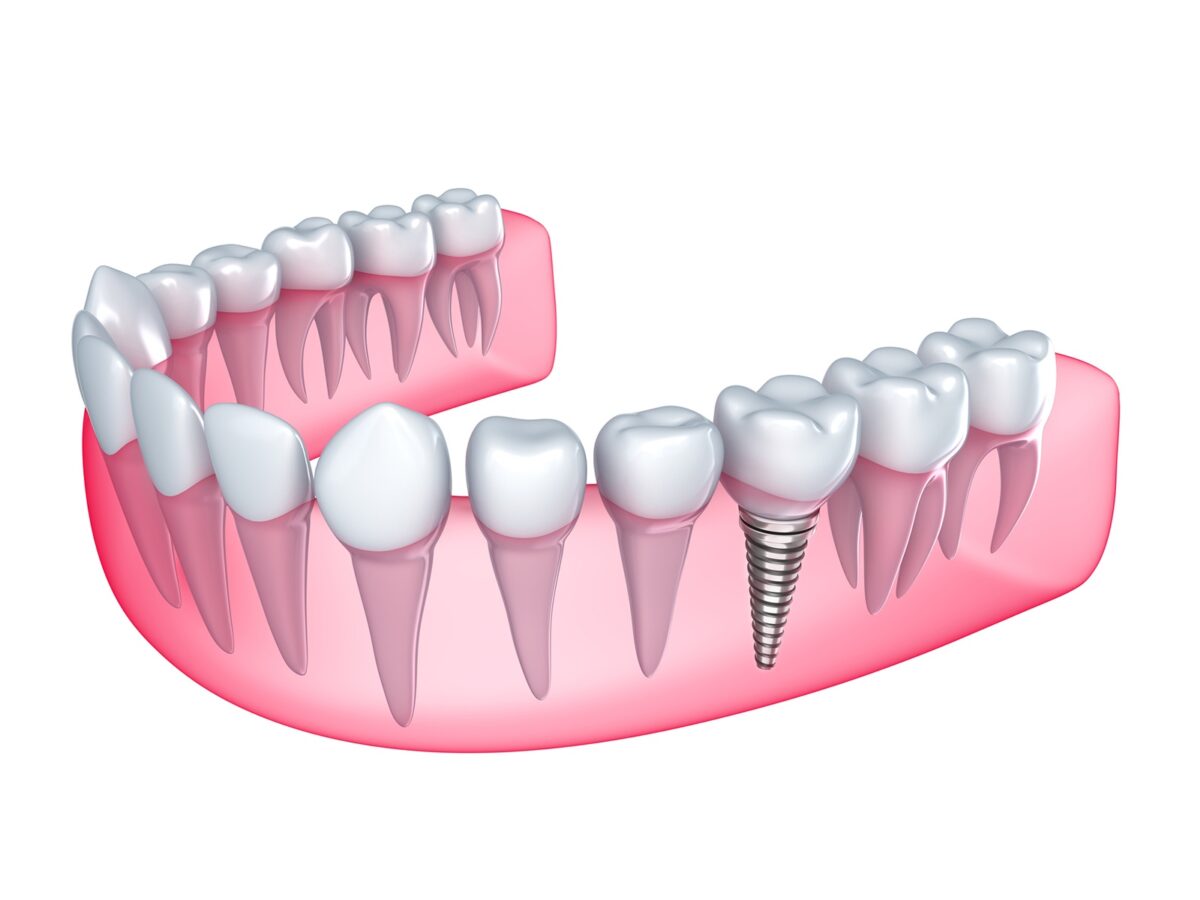
By week 6, woven bone typically bridges the gap between the host bone and the titanium surface. While this provides initial biological stability, woven bone is disorganized and possesses low tensile strength. It is not until the remodeling phase (months 3 to 6) that this immature tissue is replaced by organized lamellar bone, which is aligned to resist functional stress. The definitive answer to how long after dental implants can I eat normally is contingent upon this histological milestone. Until the lamellar bone is fully established, the interface remains susceptible to micro-fractures under heavy shear forces (e.g., chewing tough meat or raw vegetables).
Factors Influencing the Remodeling Rate
The standard timelines are subject to biological variables.
Systemic and Local Modifiers
The rate of osseointegration is not uniform across all patients. Systemic factors such as diabetes mellitus, osteoporosis, or smoking status can significantly retard angiogenesis and osteoblast activity. Locally, the density of the bone (Type 1 cortical vs. Type 4 trabecular) affects the initial stability and the speed of remodeling. In softer maxillary bone, the timeline for how long after dental implants can I eat normally is often extended by 4 to 8 weeks compared to the dense mandibular bone. The clinician must adjust the dietary protocol based on radiographic evidence of bone density and the patient’s metabolic profile.
The restoration of a normal diet is the functional endpoint of implant therapy, but it cannot be rushed. It is a biological event, not merely a healing of the soft tissue. By adhering to a diet that respects the limitations of woven bone and the dangers of micromotion, the patient ensures that the osseointegration process proceeds without interruption.