Biomaterial Integration and the Efficacy of a Composite Tooth Filling
Within the expansive clinical archives maintained by Luxe Smile Studio, the rigorous evaluation of dental biomaterials remains a paramount focus for understanding modern restorative interventions. The shift from metallic alloys to resin-based materials demands an exhaustive comprehension of organic chemistry, cellular biology, and hard tissue histomorphology. When a patient receives a composite tooth filling, they are not merely undergoing a mechanical plugging of a cavitated lesion; rather, they are the recipients of a highly complex biochemical integration between synthetic polymers and human biological substrates. This analytical discourse explores the profound, evidence-based mechanisms that govern the adhesion, polymerization, and ultimate clinical success of resin-based restorative therapies within the dynamic oral environment.
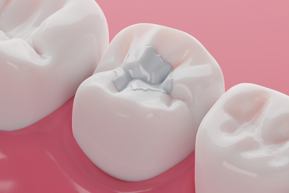
The fundamental premise of contemporary cariology dictates that the removal of infected dentin must be followed by a restoration that hermetically seals the remaining vital tissues from bacterial microleakage. A white composite dental fillings achieves this not through macroscopic mechanical retention, as was historically required by silver amalgam, but through precise micromechanical hybridization. This process begins with the deliberate chemical alteration of the enamel and dentin topography utilizing mild acidic conditioners. Phosphoric acid, typically applied at a concentration of thirty-seven percent, selectively dissolves the hydroxyapatite crystals within the enamel rods. This controlled demineralization creates a profoundly porous microscopic landscape characterized by high surface energy, which is exquisitely receptive to the capillary action of unfilled liquid resins.
Histological Adhesion Mechanisms of a Composite Tooth Filling
The true analytical complexity arises when the adhesive protocol reaches the dentinal substrate, a living tissue inherently vastly different from the highly mineralized, acellular enamel. Dentin contains a significant proportion of water and an intricate matrix of Type I collagen fibrils. When the acidic conditioner is applied to the dentin, it completely removes the smear layer—a slurry of cutting debris created during cavity preparation—and uncovers the dentinal tubules. More importantly, it demineralizes the intertubular dentin, exposing a delicate, sponge-like network of collagen fibrils. The clinical objective during the placement of a composite tooth filling is to infiltrate this exposed collagen network with hydrophilic resin monomers before the delicate fibrils collapse upon themselves.
This critical zone of integration is clinically defined as the hybrid layer. The successful formation of this layer is the singular biomechanical event that dictates the retention and marginal seal of the restoration. If the clinician desiccates the dentin too aggressively prior to resin application, the collagen network collapses, preventing the resin from penetrating. Conversely, if excessive moisture remains, the hydrophobic components of the adhesive system undergo phase separation, resulting in areas of nanoleakage. When the resin perfectly permeates the collagenous web and is subsequently polymerized via specific wavelengths of visible blue light, it forms an acid-resistant, resin-dentin interdiffusion zone that structurally links the synthetic composite material to the underlying vital tooth structure. The American Dental Association consistently highlights the mastery of this hybridization process as the cornerstone of restorative longevity.
Polymerization Shrinkage and Stress Distribution Factors
Once the adhesive layer is established, the clinician introduces the bulk restorative material. The organic matrix of this material predominantly consists of dimethacrylate monomers, such as bisphenol A-glycidyl methacrylate or urethane dimethacrylate. The transformation of this viscous paste into a rigid, functional solid is initiated by a photo-initiator, typically camphorquinone, which absorbs blue light and generates free radicals. These free radicals initiate an addition polymerization reaction, causing the individual monomer molecules to cross-link into a dense, three-dimensional polymer network.
However, this chemical conversion is accompanied by an unavoidable volumetric reduction known as polymerization shrinkage. As the monomers bind together, the distance between them decreases, causing the entire mass of the restoration to shrink by a factor of one to three percent by volume. During the placement of a composite tooth filling, this shrinkage generates immense internal stresses that are directly transferred to the bonded tooth interfaces. If the stress generated by the shrinking polymer exceeds the shear bond strength of the hybrid layer, the adhesive interface will rupture. This catastrophic failure manifests clinically as a microscopic gap at the cavity margin, immediately rendering the tooth susceptible to bacterial ingress, recurrent carious lesions, and profound postoperative hypersensitivity.
Enamel and Dentin Hybridization Protocols for a Composite Tooth Filling
To mitigate the deleterious effects of polymerization shrinkage stress, clinical analysts advocate for specific application methodologies based on robust biomechanical evidence. The incremental layering technique is universally recognized as a mandatory protocol. By applying and curing the restorative material in oblique increments no thicker than two millimeters, the clinician drastically reduces the configuration factor, commonly referred to as the C-factor, of the preparation. This controlled geometry minimizes the ratio of bonded to unbonded surfaces, allowing the restorative material to undergo free volumetric shrinkage toward the unbonded surfaces rather than pulling forcefully against the cavity walls.
Furthermore, the integration of flowable composite liners at the cervical margins of proximal boxes acts as a stress-absorbing elastic buffer. Because these low-viscosity materials possess a lower modulus of elasticity than heavily filled packable composites, they can flex microscopically during the polymerization phase of the overlying layers, effectively dissipating the internal stresses and preserving the integrity of the crucial dentinal seal.
The successful execution of a composite tooth filling demands an uncompromising adherence to biochemical principles and material science protocols. The integration of a synthetic polymer with human tooth structure is a fragile, technique-sensitive endeavor. By understanding the precise mechanisms of collagen hybridization and polymerization stress management, clinicians can reliably establish a durable, hermetic seal that restores both the biological function and the anatomical integrity of the compromised dentition.